Research / PhD · 2020 – 2025
Biophysical Modeling & Systems Biology
Modeling the cellular signaling perturbations that drive synaptic dysfunction in Alzheimer's disease.
1. Disrupted Ca²⁺ signaling, neurotransmitter release and plasticity in familial AD
Adeoye, Shah, Demuro, Rabson & Ullah · Cells, 2022
Dysregulated intracellular Ca²⁺ homeostasis plays a pivotal role early in AD, particularly in familial AD (FAD), but the mechanisms linking these molecular changes to functional impairment were unclear. We built a detailed computational model of intracellular Ca²⁺ signaling and exocytosis in hippocampal synapses to study how pathological Ca²⁺ release from the endoplasmic reticulum (ER) alters action-potential-driven transmission in FAD.
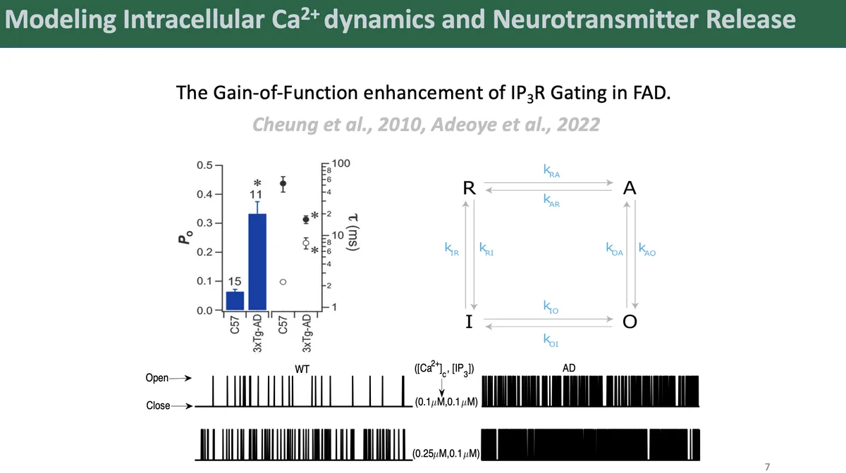
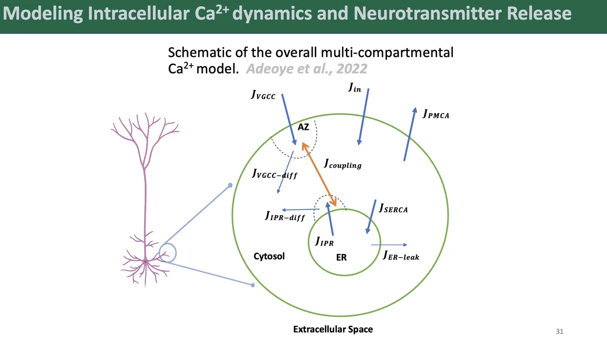
We first developed a four-state kinetic model of IP₃R gating, fit to data from cortical neurons, that captures the mechanistic basis of IP₃R gain-of-function in FAD. On top of it we built a multicompartment model of Ca²⁺ dynamics in a CA3 pyramidal axonal bouton.
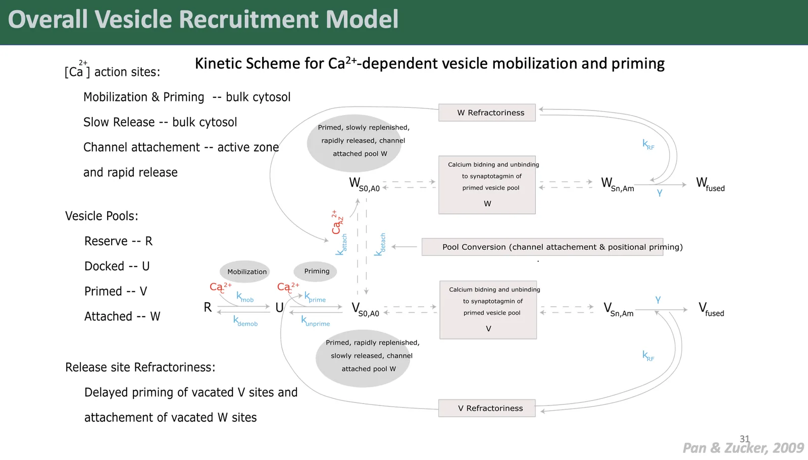
Coupling this to a detailed model of synaptic vesicle exocytosis let us examine how IP₃R-mediated ER Ca²⁺ mishandling changes release kinetics.
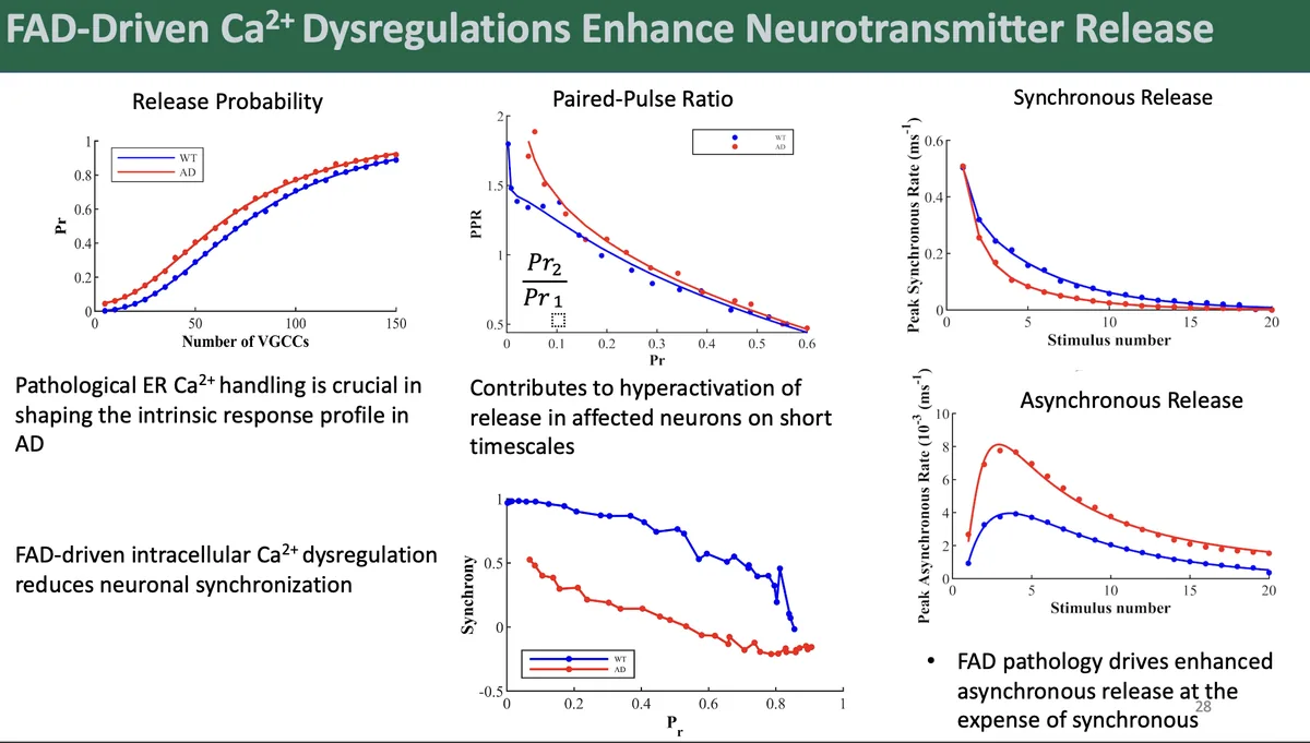
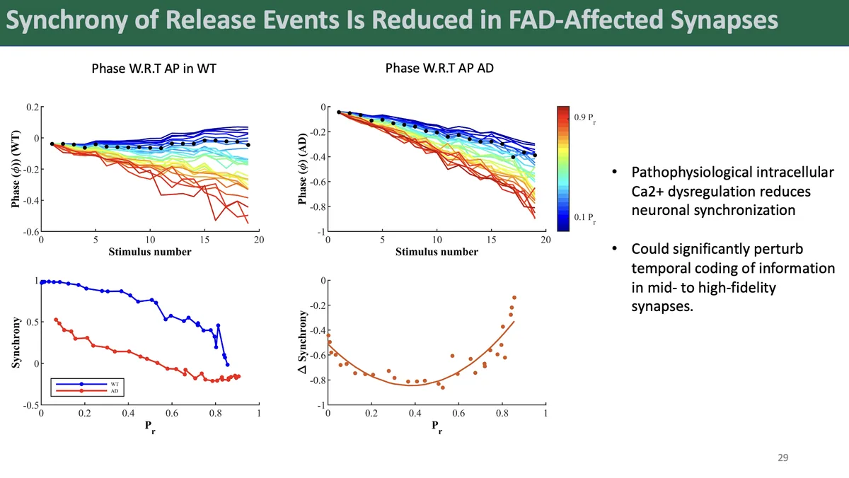
FAD synapses showed higher release rates, higher intrinsic release probability, and more vesicles released from the readily releasable pool after a single action potential, suggesting small hippocampal synapses may be most vulnerable. Contrary to the idea that high initial activity drives depression through vesicle depletion, FAD synapses showed reduced paired-pulse depression, which we attribute to sustained elevated residual Ca²⁺.
Most strikingly, the model revealed a shift toward asynchronous release at the expense of synchronous release, and impaired response synchronization, which could disrupt temporal coding in mid- to high-fidelity synapses.
2. Amyloid-β cation pores and presynaptic function
This work extends the model to Aβ-mediated cation pore formation. Oligomeric Aβ forms Ca²⁺-permeable pores in neuronal membranes, but how these pores modulate presynaptic function and release is poorly understood.
We added a nanodomain representing Aβ pore-driven Ca²⁺ transients to the CA3–CA1 synapse model, capturing localized Ca²⁺ dynamics from spatially distributed pore clusters, with gating properties (sub-conductance levels and temporal evolution) based on single-channel imaging data.
Preliminary results show prolonged release phases and altered Ca²⁺ handling, with release events redistributed in time rather than delivered in precisely timed bursts. Aβ pores play a biphasic role in synaptic activation that depends on their activity pattern, and their interaction with FAD-driven ER Ca²⁺ release appears to disrupt transmission more severely than either factor alone.