Research / PhD · 2021 – 2025
Functional Genomics & Network Biology
Characterizing the cell-type-specific molecular perturbations that drive dysregulation of biological processes in Alzheimer's disease.
1. Cell-type-specific network perturbations driving dysregulated biology in AD
Adeoye, Shah & Ullah · Aging and Disease, 2024
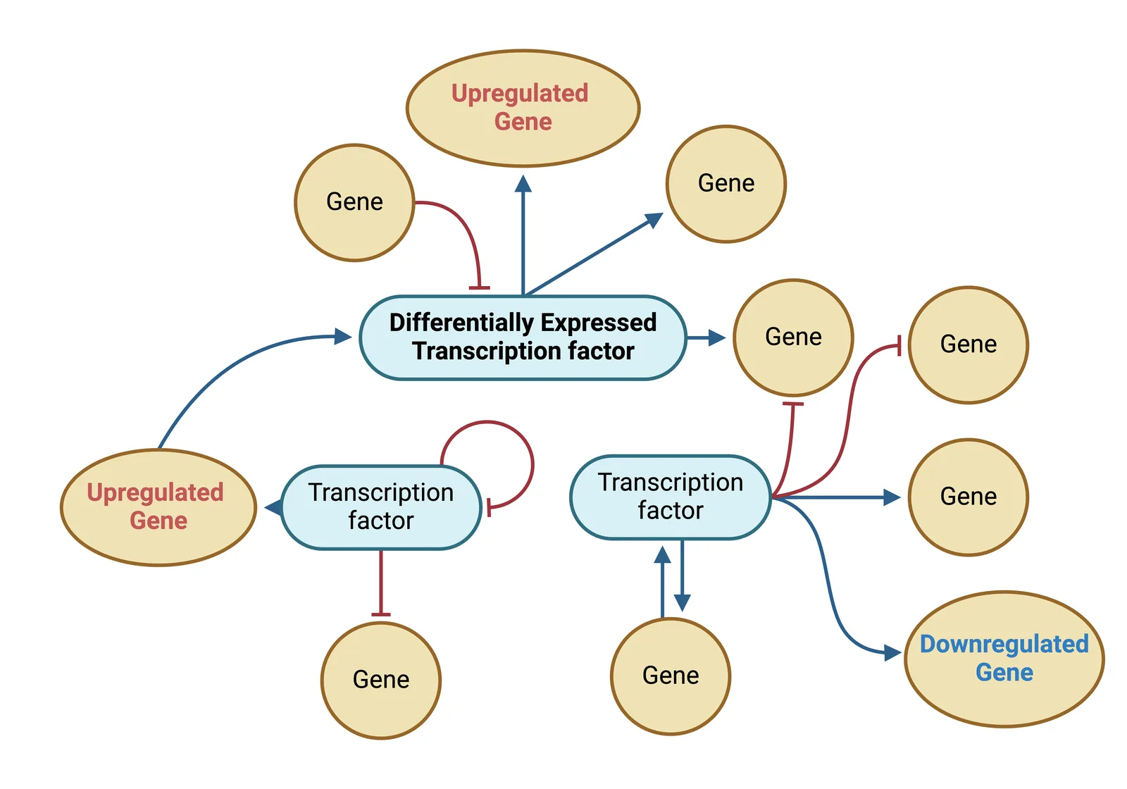
AD is a complex systems disease: its pathology extends beyond isolated molecular changes to interactions within gene networks. Transcriptomic profiling has opened up cell-type-specific questions, but traditional differential gene expression (DEG) analysis misses much of the disrupted biology. We developed a systems-biology approach to uncover the cell-type-specific drivers of biological process perturbations in AD.
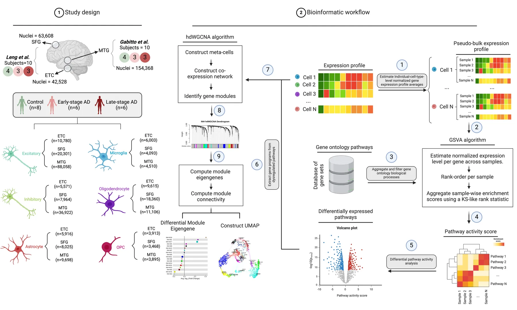
Using snRNA-seq from key AD brain regions, we combined differential activity analysis of Gene Ontology biological processes with high-dimensional weighted gene co-expression analysis (hdWGCNA). Aggregating expression within each GO process into pathway activity scores per donor, brain region and cell type increased sensitivity to subtle expression changes.
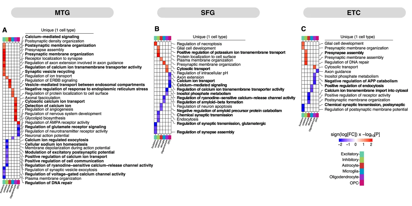
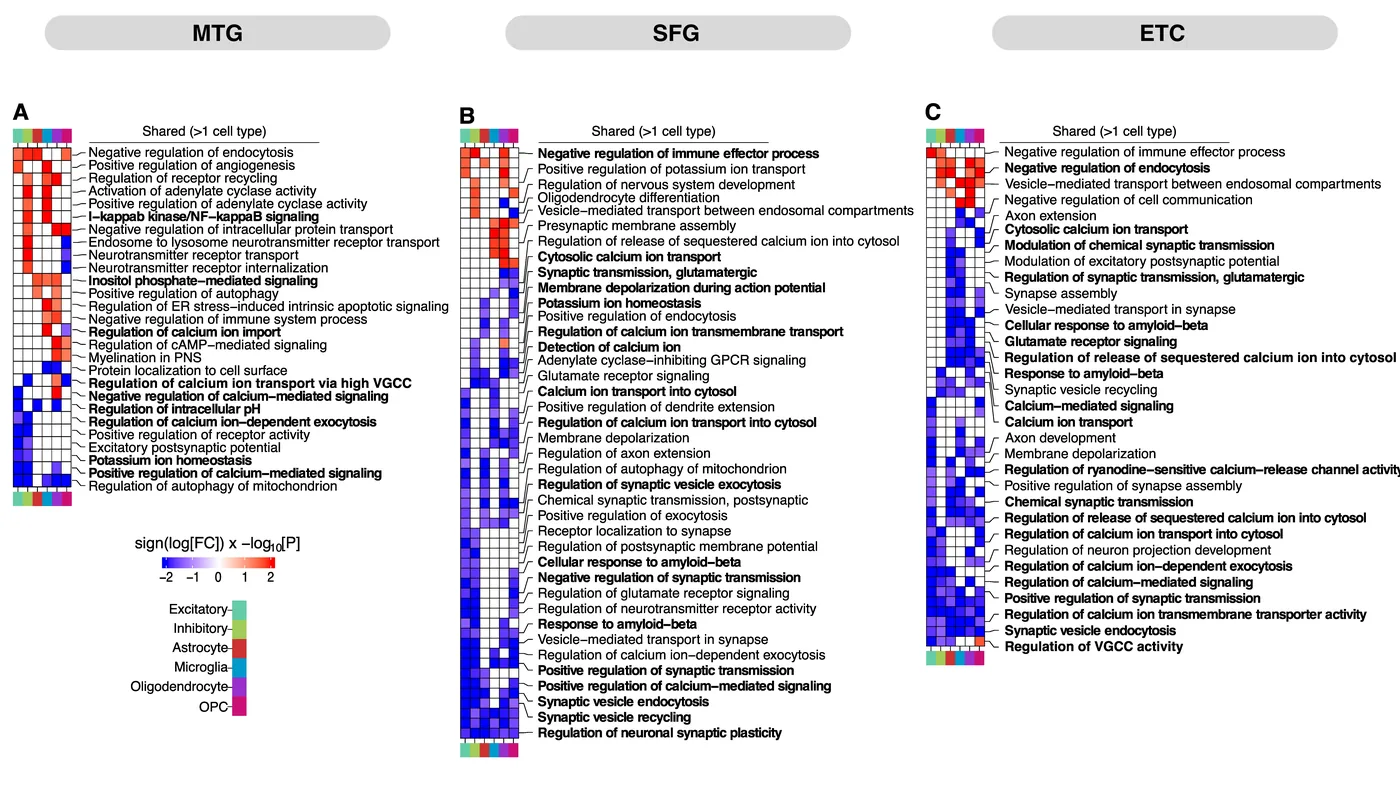
AD inflicts widespread perturbations across all three regions, from cell-type-specific alterations to pathways disrupted in many cell types. In the middle temporal gyrus, for example, neurons showed distinct changes in membrane organization, vesicle recycling and Ca²⁺ signaling, while glia predominantly showed altered vesicle transport, inflammation and signaling.
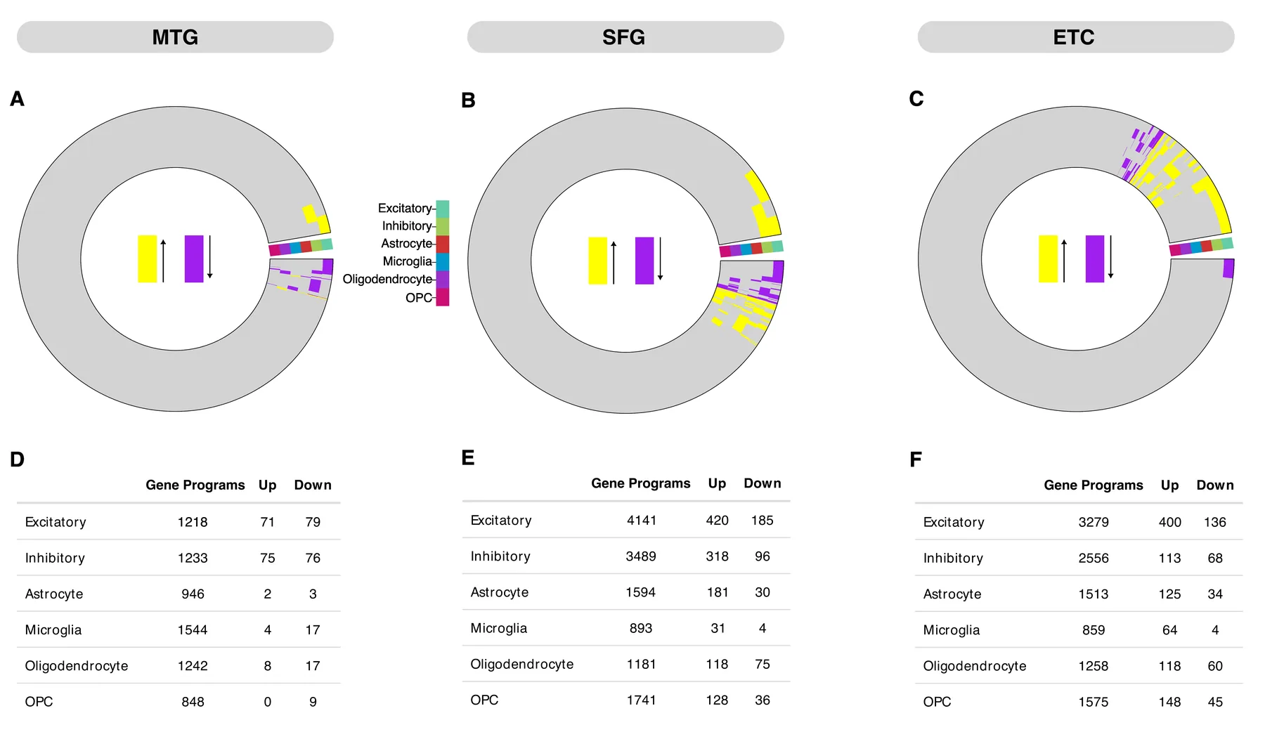
Surprisingly, only a small fraction of DEGs belonged to the gene programs that make up the perturbed pathways. Differential expression alone does not capture the complexity of AD molecular changes.
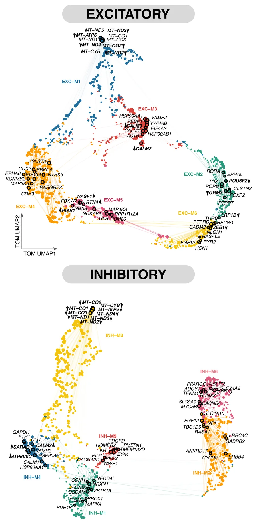
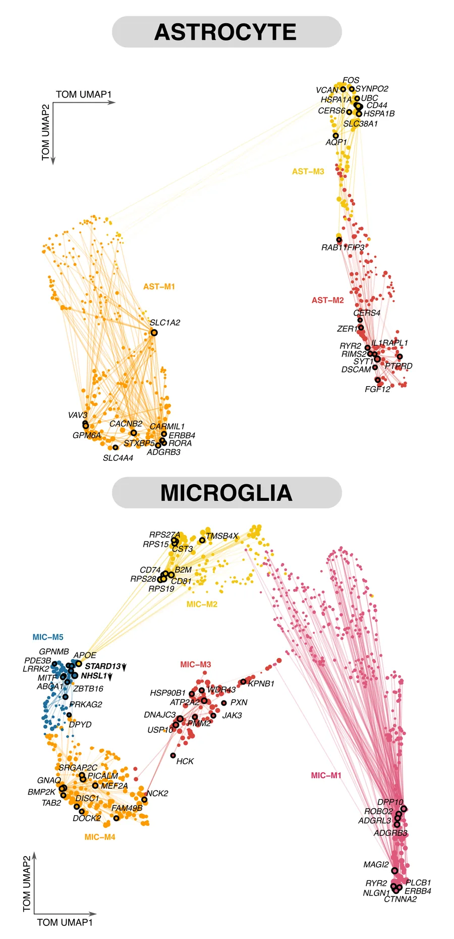
To see how DEGs influence systems-level dysregulation, we built co-expression networks per cell type and region, focused on genes within perturbed pathways. We identified distinct modules and hub genes for each cell type. In the MTG, differentially expressed hubs such as MT-CO1, CALM2 and WASF1 concentrated in specific modules, pointing to dysregulated mitochondrial function, Ca²⁺ signaling and cytoskeletal dynamics. Few glial modules were enriched for hub-DEGs, but many contained AD-related hubs such as APOE, CLU, CST3, CD81 and SLC-family genes tied to glutamate regulation, synaptic plasticity and microglial activation.
Recurrent hub genes across regions suggested conserved drivers. Excitatory neurons shared hubs such as ACTB, CALM1/2, GAPDH, HSP90AB1 and UCHL1, involved in Ca²⁺ regulation, autophagy and synaptic function. Most of these were not DEGs, reinforcing that coordinated network-level dysregulation, rather than changes in individual genes, mediates pathway perturbation.
2. Glia–neuron signaling that propagates AD pathway dysregulation
Building on these findings, this work maps how glia–neuron signaling propagates AD-associated pathway dysregulation, with a focus on astrocyte–glutamatergic neuron signaling at tripartite synapses.
Astrocytes and microglia play critical roles in neuronal dysfunction in AD, and our earlier work showed reactive signatures and perturbed regulatory programs in both. These perturbations could propagate molecular alterations from glia to neurons, contributing to synaptic dysfunction and neuronal loss, but the mechanisms, and how much is cell-autonomous versus non-cell-autonomous, remain poorly defined.
We combine scRNA-seq, scATAC-seq and complementary systems-level strategies to separate pathway disruptions that originate within a cell type from those caused by dysfunctional signaling between cell types, for example complement (C1q) expression in disease-associated microglia or disrupted thrombospondin release from astrocytes triggering synaptic pruning and excitotoxicity.
We then focus on tripartite synapses. Astrocytes regulate excitatory glutamate signaling and prevent excitotoxicity, but how this fails in AD is unclear. Using spatial transcriptomics from AD and control tissue across brain regions, we map DEGs, ligand–receptor pairs and dysregulated pathways at astrocyte–neuron interaction sites, testing the hypothesis that AD alters astrocytic receptors, transporters and channels in ways that break coordination with neighboring glutamatergic neurons.